
Figure 3.
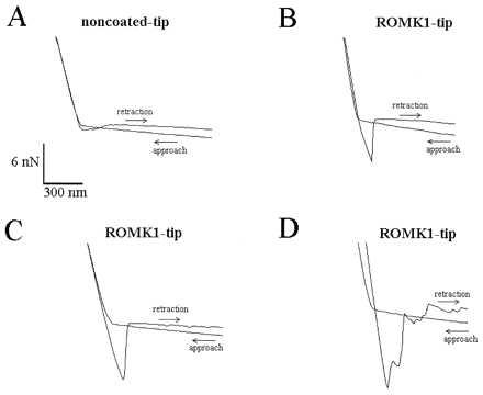
Four force scan curves illustrating the difference between noncoated AFM tips (A) and AFM tips coated with the ROMK1 channel protein (B–D). To obtain a force curve, the AFM tip first approaches the mica surface until the cantilever holding the tip is distorted. This creates the upward slope. Then, the AFM tip is retracted (downward slope) until the retractive force overcomes the adhesive force and the tip “jumps off” the mica surface. The adhesive force between a noncoated AFM tip and mica is usually small, causing a small downward-directed sink before the AFM tip jumps back into the noncontact position (A). In contrast, successfully coated tips show a significantly deeper sink before the tip retracts into the noncontact position (B–D).