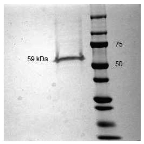
FIGURE 3. Chromatographic recombinant ERβ used in the electrophysiological experiments.
Coomassie blue staining of recombinant ERβ (Invitrogen) after SDS-PAGE reveals a single band at ~59 kDa, which corresponds to the full-length isoform ERβ1. In the right lane, bands of molecular weight standards are presented and their respective molecular weight is indicated in kDa.

