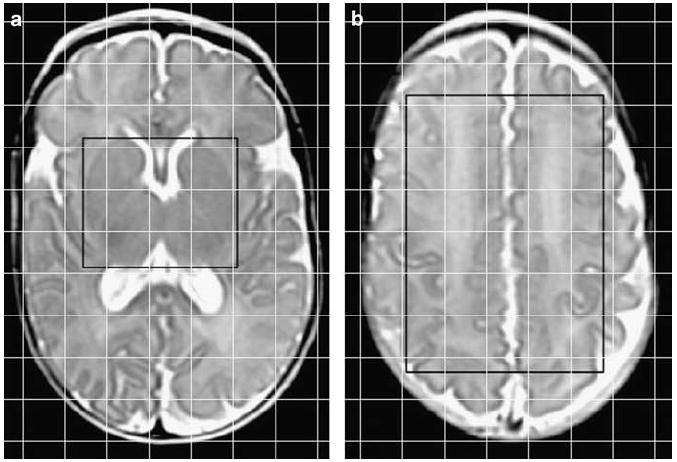
Figure 1.

Location of proton MRS regions of interest demonstrated on axial T2 images of an infant (27-week gestation) scanned at a postmenstrual age of 40 weeks. The regions of interest are within the point-resolved spectroscopy box displayed as a black rectangle, and the 2D voxel locations are displayed as a superimposed grid of white lines. (a) The region of interest at the level of the basal ganglia was comprised of the combined left and right thalamus and basal ganglia. (b) The region of interest at a supraventricular level was comprised of the cortex including tissue from the frontal, parietal and occipital lobes.
