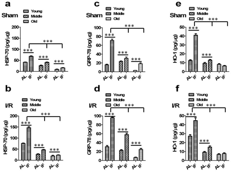
Fig 3. Levels of HSP-70, GRP-78 and HO-1 are increased in the brains of mice on the IF diet and are decreased with aging.

(a) HSP-70 quantified from brain lysates of sham animals. HSP-70 levels were significantly decreased in middle aged and old sham animals compared to young sham animals (n=10 in each group). ***p<0.0001 compared with young sham animals. IF sham animals had significantly increased HSP-70 compared to AL-fed sham controls in all age group animals. ***p<0.0001 compared with AL sham animals in each group. (b) IF I/R animals had significantly increased HSP-70 compared to AL-fed I/R controls in all age group following cerebral I/R. ***p<0.0001 compared with AL I/R animals in each group. (c) GRP-78 levels in the brain were significantly decreased in middle aged and old sham animals compared to young sham animals (n=10 in each group). ***p<0.0001 compared with young sham animals. IF sham animals had significantly increased GRP-78 compared to AL-fed sham controls in all age group animals. ***p<0.0001 compared with AL sham animals in each group. (d) Following cerebral I/R, IF animals had significantly increased GRP-78 compared to AL-fed I/R controls in all age group. ***p<0.0001 compared with AL I/R animals in each group. (e) HO-1 levels were significantly decreased in middle aged and old sham animals compared to young sham animals (n=10 in each group). ***p<0.0001 compared with young sham animals. IF sham animals had significantly increased HO-1 compared to AL-fed sham controls in young and middle age group. There was no significant increase observed in IF old animals compared to AL-fed old animals. ***p<0.0001 compared with AL sham animals. (f) IF young and middle aged I/R animals had significantly increased HO-1 compared to AL-fed I/R controls following cerebral I/R. ***p<0.0001 compared with AL I/R animals.
