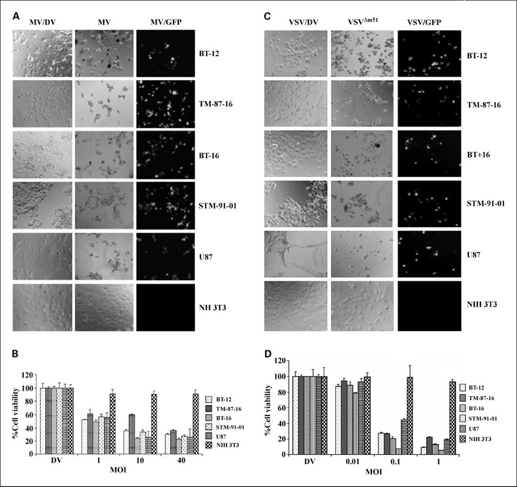
Fig. 1.
Rhabdoid tumor cell lines are susceptible to oncolysis by MV and VSVΔM51 in vitro. A, rhabdoid tumor cell lines were infected with 10 MOI of live MV or UV-inactivated dead MV (MV/DV). CPE (middle column) and expression of virally encoded GFP (right column) were evident in all malignant rhabdoid tumor cell lines 72 h after exposure to live MV (magnification, × 200). B, relative cell viability was measured using MTT assays 72 h postinfection with the indicated MOI of MV. C, tumor cell lines were infected with 1 MOI of live or UV-inactivated dead VSVΔM51 (VSV/DV). CPE (middle column) and expression of virally encoded GFP (right column) were evident in all rhabdoid tumor cell lines 72 h after exposure to live VSVΔM51 (magnification, × 200). D, relative cell viability was measured using MTT assays 72 h postinfection with the indicated MOI of VSVΔM51. U87 glioma cells and NIH 3T3 cells served as positive and negative controls, respectively.

