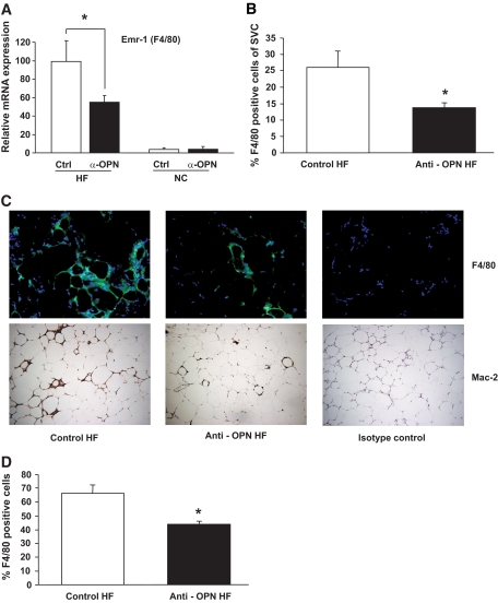
FIG. 2.
Adipose tissue macrophage accumulation is reduced by OPN neutralization. Obese high-fat diet–fed (HF) and lean normal chow–fed (NC) mice were treated with OPN-neutralizing (Anti-OPN) or control antibody (n = 8 per group for HF and n = 5 per group for NC mice). A: mRNA expression of the macrophage marker F4/80 (encoded by Emr1 gene) was analyzed in GWAT by real-time RT-PCR. The mean of control HF was set to 100%. B: Percentage of macrophages (F4/80-positive cells) in the SVC fraction of GWAT as determined by flow cytometry. C: Adipose tissue macrophage accumulation was determined by immunofluorescence of F4/80+ cells (upper row) and immunohistochemical staining of Mac-2+ cells (bottom row) in GWAT isolated from high-fat diet–fed mice after anti-OPN or control antibody treatment. Representative pictures are given in 40-fold magnification. D: Adipose tissue macrophages as detected by F4/80 positivity in tissue sections were counted as F4/80+ cells relative to total number of cells. (A high-quality digital representation of this figure is available in the online issue.)

