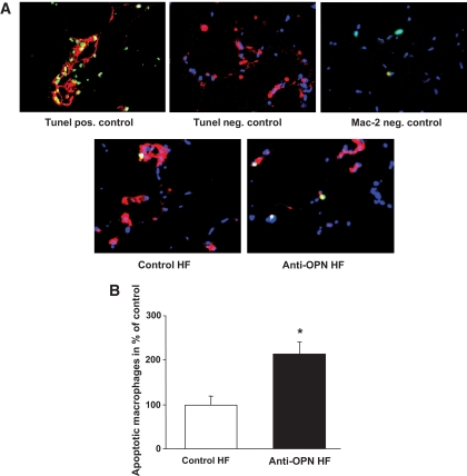
FIG. 3.
A: Apoptotic cells were determined by tunel staining (green), macrophages were stained red by immunoflourescence using anti-F4/80 monoclonal antibody on frozen sections. Representative pictures are given. B: Quantification of apoptotic macrophages (TUNEL and F4/80 double-positive cells per F4/80-positive cells). The mean of Control HF was set to 100%. *P ≤ 0.05. (A high-quality digital representation of this figure is available in the online issue.)

