
Figure 5.
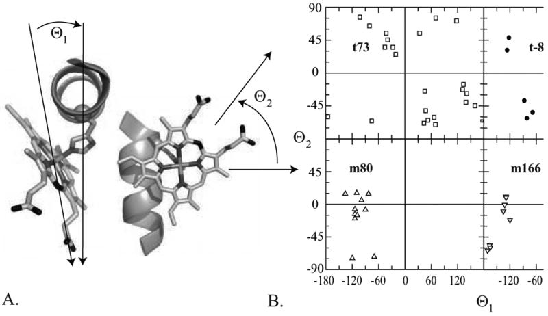
t73 rotamers have non-specific heme propionate orientations in the dataset while the other rotamers cluster at specific orientations. (A) Starting with the helix aligned upright with the N-terminus pointing at the observer, the azimuthal angle θ1 is formed between the vector normal to the helix vector which passes through the histidine C(α) and the iron-heme CHA vector. The zenith angle θ2 is measured between helical vector and the iron-heme CHA vector oriented such that both are same plane. The heme-helix model depicted has been placed at the mean θ1 and θ2 angles for the c-type cytochromes in the m166 rotamer. (B) The distribution of θ1 and θ2 angles of the hemin-containing proteins in the dataset subdivided by rotamer. In the m166 dataset, b-type hemes are depicted using open triangles and c-type hemes are depicted using solid triangles.