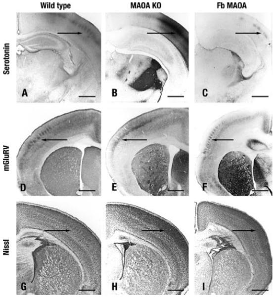
FIGURE 6. Cortical alterations in S1 of MAO-A KO and forebrain transgenic mice at postnatal day 7.

A–C, thalamocortical axon segregation visualized with 5-HT IR. A, wild type. C, forebrain (Fb) transgenic mice 5-HT IR forms whisker-related patches in layer IV of S1 (arrow). B, in MAO-A KO mice thalamocortical axons do not form patches. D–F, dendritic differentiation visualized with mGluR5 IR. D, wild type. F, in forebrain transgenic mice, mGluR5 IR labels barrels in layer IV (arrow), whereas they do not form in MAO-A KO mice (E). G–I, cytoarchitectonic differentiation. G, in wild type mice granular neurons form barrels in layer IV (arrow). H, MAO-A KO. I, forebrain transgenic mice granular neurons do not form barrels (arrow). Scale bars = 560 μm.
