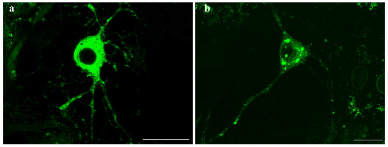
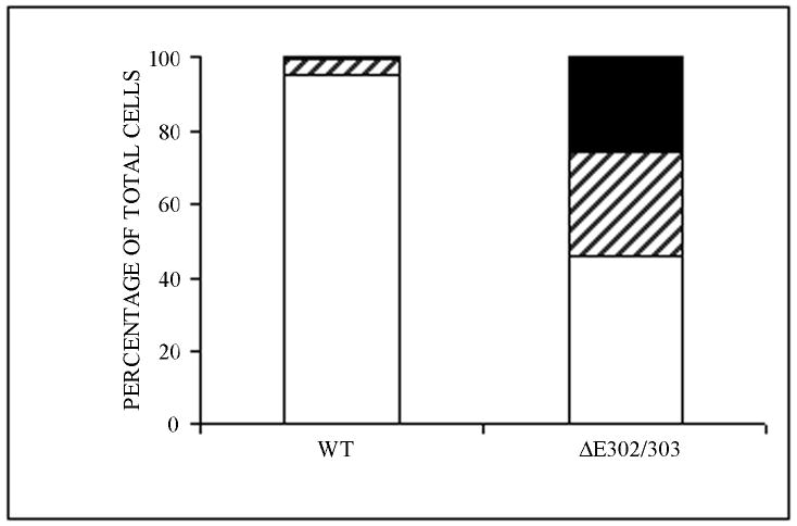
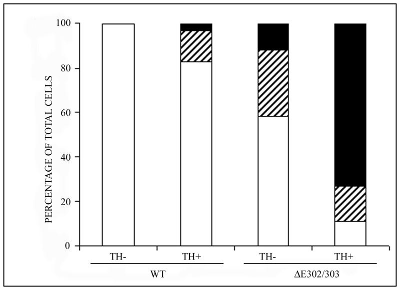
Figure 3.
Mutant torsinA promotes inclusion formation in catecholaminergic neurons. Primary midbrain cultures were transduced with HSV vectors expressing wildtype (a) or ΔE302/303 (b) mutant torsinA and stained with polyclonal anti-torsinA and anti-rabbit AlexaFluor 488. Scale bar = 20μM. C. For each experiment we counted the number of transduced cells and calculated the number of cells that had no inclusions (open bar), a few small aggregates (<1μM, hatched bar) or one or more large inclusions (>1μM, heavy shaded bar). Quantification demonstrated a significant difference in the number of cells showing inclusion formation after transduction with either the wildtype or mutant torsinA construct (wildtpe: 5±0.9% Vs ΔE302/303: 53.7±3.6%, mean±SD, p<0.0001, n=2050 neurons counted). D. Inclusion formation was influenced by neuronal subtype. Analysis as in C demonstrated wildtype torsinA did not generate inclusions in the TH- neurons but inclusions were seen in approximately 20% of TH+ neurons. In contrast, mutant torsinA formed inclusions in approximately 40% of TH- neurons but over 80% of TH+ neurons (TH-: 42.8±3.8% Vs TH+: 84.2±1.4%, mean±SD, p<0.0001, n=1140 neurons counted). Mutant torsinA formed significantly more inclusions in TH+ neurons compared to wildtype (wildtype: 20.2±8.8% Vs ΔE302/303: 84.2±1.4%, mean±SD, p=0.0002, n=567 neurons counted).


