
Figure 6. Scanning electron microscopic analysis of tetherin interaction with budding virions and models for tetherin incorporation.
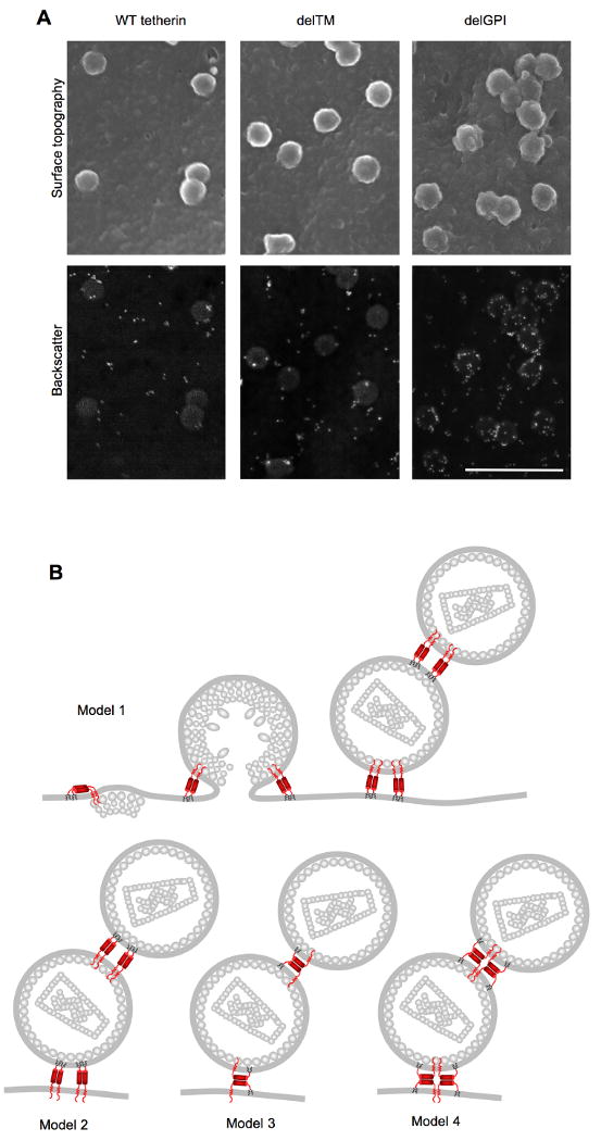
(A) Scanning electron microscopic analysis of 293T cells transiently expressing HA-tagged WT, delTM or delGPI tetherin proteins and HIV-1 Gag bearing mutations in the PTAP L-domain sequence. Cells were fixed and stained with anti-HA primary antibody and anti-mouse IgG-gold conjugate. Surface topography (upper panels) reveals virion particles, while backscatter electron detection (lower panels) reveals gold particles (white) marking the position of tetherin molecules. Scale bar = 500nm
(B) Models for incorporation and virion tethering by the tetherin protein. Several stages of HIV-1 assembly are depicted for model 1, and only tethered virions are shown for models 2-4. In model 1, the TM domains of a tetherin dimer are incorporated into the virion envelope, and the GPI anchors remain embedded in the host-cell membrane. In model 2, the reverse situation occurs. In model 3, only one of a pair of disulfide linked tetherin molecules has both membrane anchors incorporated into the virion envelope. In model 4, one disulfide linked tetherin dimer incorporated into the virion envelope interacts with another dimer in the host-cell membrane via coiled-coil based interactions.