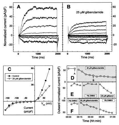
Figure 2.

Partial inhibition by glibenclamide of outward potassium channels in V. faba guard cell. (A) Recordings of normalized inward (seen as downward deflections) and outward (upward deflections) potassium currents in whole-cell configuration. (B) Thirty minutes after perfusion with 25 μM glibenclamide, outward but not inward potassium currents were partially inhibited. (C) Superposition of current–voltage curves of the cell in A before (•) and after bath perfusion (B) with 25 μM glibenclamide (▿). (D) Reduction in the steady-state normalized outward potassium current vs. time after glibenclamide perfusion. Values correspond to the experiment described in A–C. Upward pointing arrows refer to current recordings illustrated in A and B. (E) Typical experiment illustrating the time course of the normalized outward potassium current. Channel activity was unaffected by DMSO perfusion but still sensitive to glibenclamide inhibition. (F) Even after a 20-min glibenclamide wash-out, the normalized outward potassium current was not recovered. (A–D) Whole-cell capacitance was 10 pF. Seal resistance was 2.5 GΩ. (D–F) Time course of the outward K+ current is given for a membrane potential of +80 mV. Changes of the bath solution are represented by alternating open and shaded areas.
