
Fig. 6.
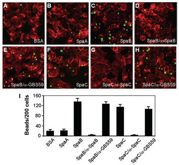
SpaB- or SpaC-mediated adherence of latex beads to pharyngeal cells.
Semi-confluent cells grown on coverslips were infected with protein-coated beads for 1 h.
The washed cells were fixed and stained with Texas-Red-X phalloidin.
A–H. Shown here are the fluorescence images of cells incubated with fluorescent beads bound to BSA (A), SpaA (B), SpaB (C), SpaB blocked with α-SpaB (D), SpaC (F) and SpaC blocked with α-SpaC (G). Beads with bound protein incubated with an unrelated antibody against a pilin of Streptococcus agalactiae, α-GBS59, were used as controls (E and H).
I. The results are shown as an average of the numbers of beads bound per 200 cells of three independent experiments (with standard deviation ± SD).