Fig. 3.
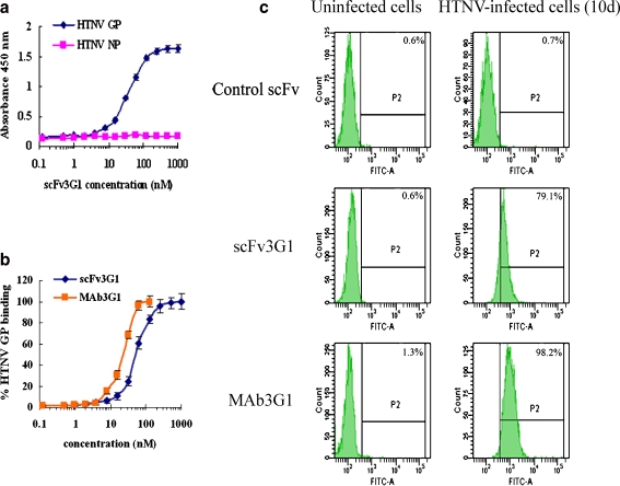
HTNV GP antigen-specific binding characteristics of scFv3G1. a Solid-phase binding of purified scFv3G1 to HTNV GP antigen as measured by ELISA. The wells were coated with HTNV GP or HTNV NP and then incubated with purified twofold serially diluted scFv3G1 from 1,000 to 0.1 nM. b To compare the HTNV GP-specific antigen-binding activity of scFv3G1 to that of MAb3G1, serially diluted scFv3G1 and MAb3G1 were assayed. Results are plotted as percentages of HTNV GP binding. c Flow cytometric analysis to evaluate whether scFv3G1 can bind HTNV GP antigen expressed on the cell membrane. Ten-day HTNV-infected Vero E6 cells or uninfected cells were incubated with 100 nM purified scFv3G1 and followed by detection with FITC-conjugated anti-His-Tag antibody

