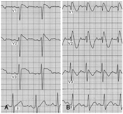
Fig. 4.
ECG changes of patients in case 2 after the flecainide challenge test. A: ECG of a 12 year old boy after the flecainide challenge test. Typical coved ST-segment elevation followed by T wave inversion is observed on V1 and V2 leads. B: ECG of a 10 year old girl after the flecainide challenge test. Note the typical Brugada pattern ST-segment elevation on V1 and V2 leads. ECG: electrocardiogram.

