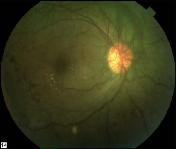
Figure 2. Advanced proliferative diabetic retinopathy with neovascularization of the disc.
Scattered retinal hemorrhages, macular exudates, and a cotton-wool spot are present. This fundus image of the right eye appears green because of the increased permeability of the abnormal vessels to fluorescein following an angiogram.

