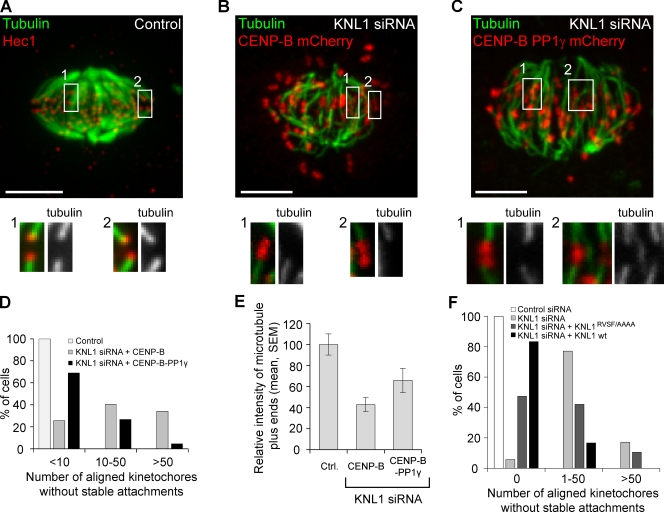
Figure 4.
PP1 at kinetochores stabilizes microtubule attachments. HeLa cells were treated as indicated, fixed, and analyzed for cold-stable microtubules. (A–E) Cells were transfected with KNL1 siRNA and either CENP-B–PP1γ-mCherry to target exogenous PP1 to kinetochores, or CENP-B–mCherry as a control. An untransfected control cell (A) was analyzed the same way and stained for Hec1 to label kinetochores. Images (A–C) are maximum intensity projections of confocal stacks; insets show enlarged views (indicated by the numbered boxed regions) of optical sections showing individual kinetochores. Bars, 5 µm. (D) Cells were classified by the number of aligned kinetochores lacking cold-stable microtubule fibers (n ≥ 45 cells in each group). (E) The intensity of microtubule plus ends at attached kinetochores (n > 60 from multiple cells) was measured in each condition. (F) Cells were transfected with KNL1 siRNA and either siRNA-resistant wild-type KNL1 or the KNL1RVSF/AAAA mutant (n ≥ 25 cells in each group), and analyzed as in D. Note that the binning in D and F is different because the effect of mutating the RVSF motif is more subtle than the effect of depleting KNL1.

