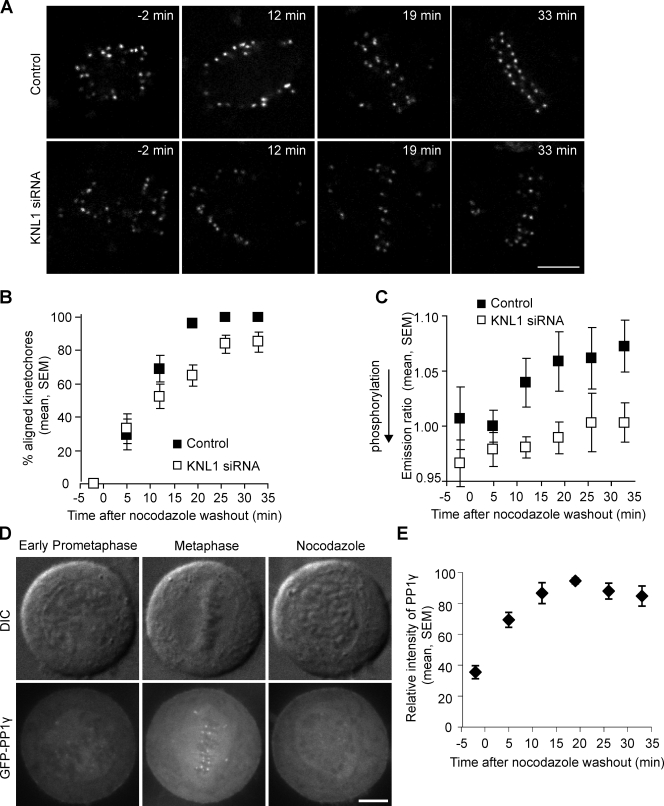
Figure 5.
PP1γ is recruited to kinetochores and dephosphorylates an Aurora B substrate as centromere tension is established. (A–C) HeLa cells transfected with a kinetochore-targeted Aurora B phosphorylation sensor, with or without KNL1 siRNA, were imaged live during recovery from nocodazole (30 ng/ml). Cells were followed for 33 min, which was sufficient time for control cells to reach metaphase. Cells rarely entered anaphase during this time, and any anaphase cells were excluded from the analysis. Representative images (A) show YFP emission. At each time point, the percentage of kinetochores aligned at the metaphase plate was determined (B) and the YFP/TFP emission ratio was calculated (C). Each data point represents seven cells, >15 kinetochores per cell. (D) Images of HeLa cells stably expressing GFPLAP-PP1γ in early prometaphase, metaphase, or treated with nocodazole. (E) The relative intensity of GFP-PP1γ at kinetochores was calculated during recovery from nocodazole (30 ng/ml). n = 6 cells, multiple kinetochores per cell. Bars, 5 µm.

