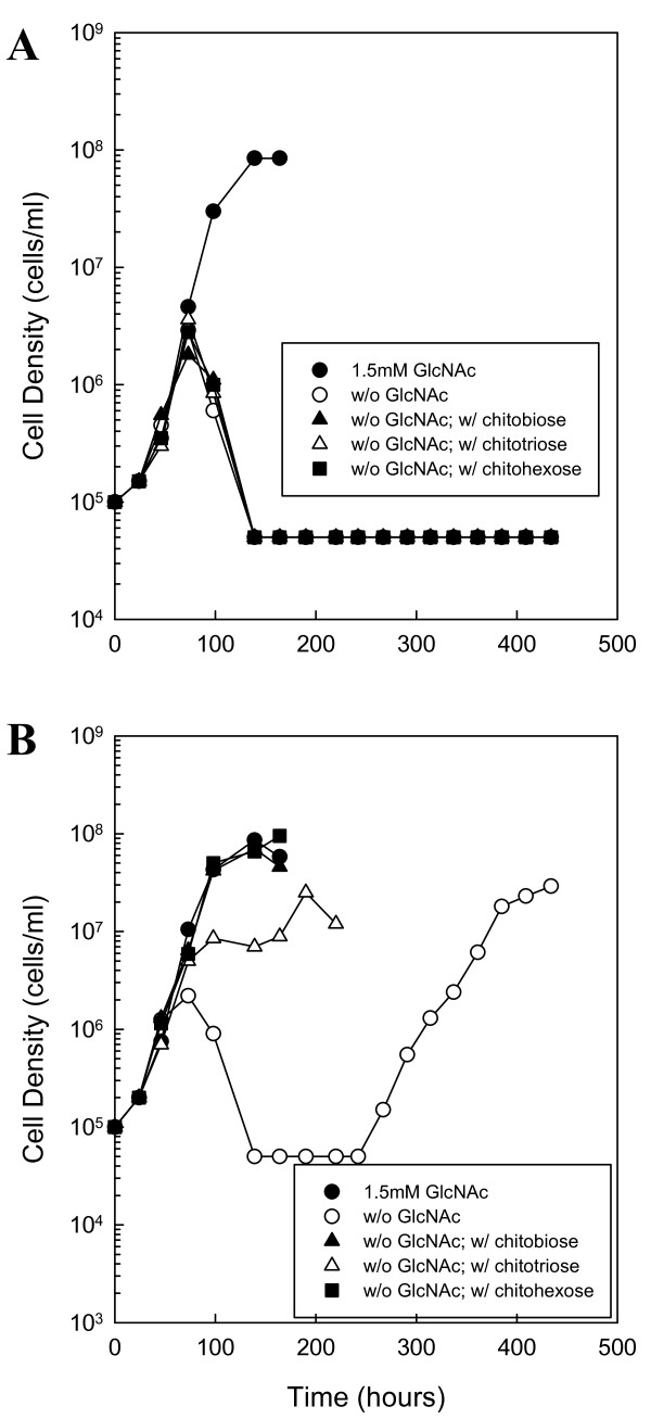
Figure 5.
Growth of a chbC mutant and complemented mutant on chitin. (A) Growth of RR34 (chbC mutant) in the presence of chitobiose, chitotriose and chitohexose. Late-log phase cells were diluted to 1.0 × 10⁵ cells ml-1 in BSK-II containing 7% boiled serum, lacking GlcNAc and supplemented with the following substrates: 1.5 mM GlcNAc (closed circle), No addition (open circle), 75 μM chitobiose (closed triangle), 50 μM chitotriose (open triangle) or 25 μM chitohexose (closed square). Cells were enumerated daily by darkfield microscopy. (B) Growth of JR14 (RR34 complemented with BBB04/pCE320) in the presence of chitobiose, chitotriose and chitohexose. Late-log phase cells were diluted to 1.0 × 10⁵ cells ml-1 in BSK-II containing 7% boiled serum, lacking GlcNAc and supplemented with the following substrates: 1.5 mM GlcNAc (closed circle), No addition (open circle), 75 μM chitobiose (closed triangle), 50 μM chitotriose (open triangle) or 25 μM chitohexose (closed square). Cells were enumerated daily by darkfield microscopy. These are representative growth experiments that were repeated four times.

