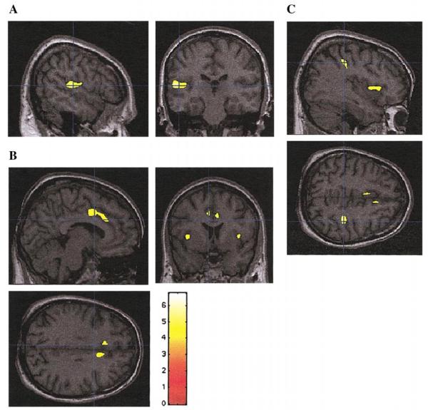
FIG. 3.
SPM{t} statistically significant regions (using spatial extent as in Table 1 and Fig. 2) superimposed on selected sections of a spatially normalized brain from a control subject. (A) Left STG; (B) left and right insula and anterior cingulate gyri; (C) Right parietal lobe, inferior parietal lobule (and insula). Neurological convention as in Fig. 1.

