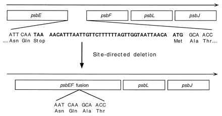
Figure 1.

Schematic diagram illustrating how the α and β subunits of cytochrome b559 were fused by connecting the psbE (α subunit) and psbF (β subunit) genes in Synechocystis 6803. Site-directed deletion of the 42 nucleotides shown in boldface type, which includes the stop codon of the psbE and the start codon of the psbF genes, resulted in the T363 mutant strain in which the psbE and psbF genes are translationally fused together.
