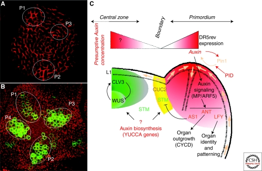
Figure 2.
From dynamic transport to patterning: Auxin and organogenesis at the shoot apical meristem of Arabidopsis thaliana. (A) Immunodetection of PIN1 efflux carrier in the L1 (top view). The image was obtained by confocal microscopy. Note the subcellular polarized localization of the auxin transporter in most cells. The localization suggests that auxin accumulates in these young organ primordia (named P1, P2, and P3 from the oldest to the youngest organ). Adapted from de Reuille et al. (2006). (B) Expression of the synthetic DR5rev::GFP reporter in the inflorescence meristem (top view). Projection of serial optical sections obtained by confocal microscopy. The green corresponds to the GFP and the red to the autofluorescence of meristematic cells. DR5 expression in young emerging primordia indicates activation of auxin induced-genes. (C) Schematic representation of the role of auxin during organogenesis in the inflorescence meristem of Arabidopsis thaliana. See text for details.

