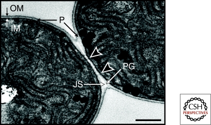
Figure 3.
Transmission electron micrograph of the junction between two vegetative cells. Arrowheads indicate microplasmodesmata, which are potential cell-to-cell channels. Note the "junctional space" between the cell wall peptidoglycan layers of the two cells, which may indicate a partial barrier between the periplasmic compartments of adjacent cells. IM, inner membrane; OM, outer membrane; P, periplasm; PG, peptidoglycan cell wall; JS, junctional space. Size bar, 0.5 µm.

