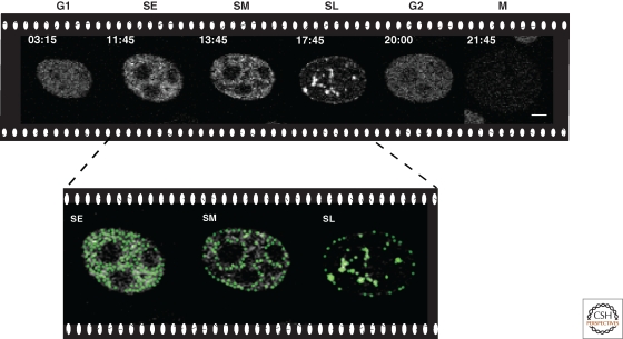
Figure 2.
Temporal progression of genome replication. Snapshots of a time-lapse confocal microscopy movie of DNA replication throughout the cell cycle in human HeLa cells stably expressing GFP-tagged PCNA. Times are indicated in hours:minutes and cell cycle phases as G1/SE (S early)/SM (S mid)/SL (S late)/G2/M. Scale bar, 5 µm. During early S phase, small foci distributed throughout the nucleus and mostly corresponding to euchromatic genomic regions are duplicated. Subsequently, the DNA replication machinery loads at perinuclear and perinucleolar heterochromatin regions followed at later times by large constitutive heterochromatic chromosomal regions. This temporal replication program is recapitulated at each cell cycle. See also Movie 1 online at http://cshperspectives.cshlp.org/.

