Figure 2.
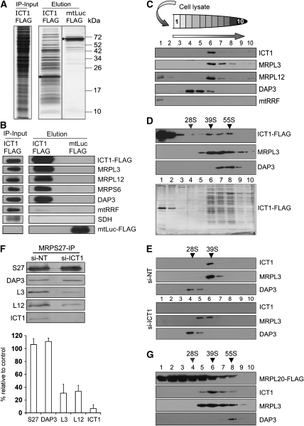
ICT1 is an integral component of the mitoribosome. (A, B) FLAG-tagged ICT1 immunoprecipitates mitoribosomes. HEK293T cells expressing FLAG-tagged ICT1 or mitochondrially localised luciferase (mtLuc-FLAG) were induced for 3 days; mitochondria were isolated, lysed and subjected to IP as detailed. The eluate and mitochondrial lysate before IP (IP-input) were separated by 15% SDS–PAGE and visualised by silver staining. * designates the FLAG protein. (B) Aliquots of the eluates were also subjected to western blot analysis with the indicated antibodies: MRPL3, MRPL12, MRPS6 and DAP3 as mitoribosomal markers; mtRRF, mitoribosome recycling factor; SDH, 70-kDa component of complex-II. (C) ICT1 co-sediments with the large mitoribosomal subunit. HeLa cells were lysed (600 μg), separated through a 10–30% sucrose gradient and fractionated as detailed (HeLa and HEK293T lysates gave identical separations). Components of the 39S mt-LSU (MRPL3, MRPL12) and 28S mt-SSU (DAP3) mitoribosomal subunits were visualised by western blotting. On immediate lysis, mtRRF is used as a matrix-soluble marker. (D) ICT1 also co-sediments with the intact monosome. Mitochondria (3 mg) of ICT1-FLAG-expressing HEK293T cells were subjected to FLAG IP; the entire eluate was separated by isokinetic density gradients and fractions were blotted as detailed above or visualised by silver staining (lower panel). Mitochondrial SSU (DAP3) and mt-LSU (MRPL3) MRPs are visualised. The approximate indicators for 28S mt-SSU, 39S mt-LSU and 55S monosome are shown and were determined as described under Materials and methods. (E) ICT1 is an integral member of 39S mt-LSU. Cell lysates (600 μg) from ICT1-depleted (si-ICT1B) or non-targeted control cells (si-NT) were separated by isokinetic gradients and proteins were visualised in the fractions by western blotting as described. Sedimentation markers were identified as above. (F) Loss of ICT1 causes depletion of the monosome. Cells expressing MRPS27-FLAG were treated with si-NT or si-ICT1B, after which IP was performed. To assess monosome formation, levels of MRPL3 and MRPL12 were quantified by western blotting of three individual experiments (right panel; MRPL3 P=0.001, MRPL12 P<0.001, MRPS27 P=0.3). (G) ICT1's association with mitoribosomes is not FLAG-dependent. Mitochondria from cells expressing MRPL20-FLAG were subjected to FLAG IP and the eluate was analysed by western blotting after isokinetic density gradients as described in panel D.

