Figure 8.
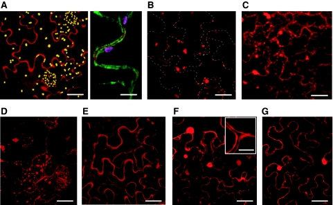
Localisation of phytaspase-mRFP in N. tabacum leaves (confocal microscopy). (A) Apoplastic localisation of phytaspase-mRFP. Agrobacterium-mediated expression of phytaspase-mRFP (48 hpi) results in largely apoplastic fluorescence (left panel) as confirmed by co-expression of this construct with the plasma membrane marker EGFP-LT16b, with clear mRFP fluorescence detected outside the cell membrane (right panel). Yellow (left panel) and purple (right panel) granules are chloroplasts showing natural autofluorescence. (B) Inhibition of phytaspase-mRFP secretion by BFA. BFA (10 μg/ml) was infiltrated 24 h after agroinfiltration of the phytaspase-mRFP construct and fluorescence was monitored 24 h later. Phytaspase-mRFP forms aggregates within the cell treated with BFA. Dashed lines show cell borders. (C) Phytaspase-mRFP is partially redistributed into cytoplasm during TMV-mediated HR. N. tabacum plants were simultaneously agroinfiltrated with phytaspase-mRFP construct and infected with TMV at 30°C. HR was induced by temperature shift to 24°C 24 hpi, and fluorescence was monitored 24 h later. (D) Redistribution of phytaspase-mRFP during stress induced by MV (similar redistribution was observed during NaCl-induced PCD). MV (10 μM) was applied 24 hpi, and fluorescence was monitored 24 h later (E) Apoplastic localisation of cathepsin B (Gilroy et al, 2007) is retained after stress treatment exemplified here with MV. Cathepsin B-mRFP fusion construct was agroinfiltrated into N. tabacum leaves and MV was applied 24 hpi. Fluorescence was monitored 24 h later. (F, G) Intracellular (presumably cytoplasmic and nuclear) localisation of free mRFP (used as an additional control) expressed from Agrobacterium in untreated N. tabacum leaves (F) was not changed during MV-induced PCD (G). Insert in (F) clearly shows unlabelled (by free mRFP) apoplastic space close to cell junctions. Of note, intracellular localisation of free mRFP (F, G) was different from that of phytaspase–mRFP under stress conditions (C, D). Scale bars are 15 μm [A-left panel, B, C, D, E, F (apart from insert) and G], 50 μm (A-right panel and insert (in F).

