Figure 7.
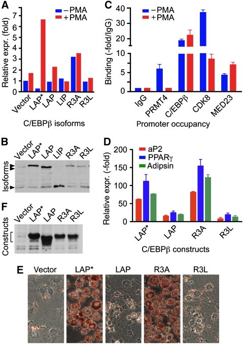
Myeloid and adipogenic gene regulation. (A) Expression of neutrophile elastase (hELA2) transcripts by C/EBPβ isoforms in NIH 3T3 cells before and after PMA treatment. NIH 3T3 cells were transfected with constructs (as indicated) and after 24 h stimulated with phorbol ester (PMA) for 1.5 h. Total mRNA and cDNA were prepared and hELA2 gene expression was analysed by RT–PCR and normalized to GAPDH expression. (B) Expression control of C/EBPβ isoforms detected by immunoblotting. (C) ChIP assay from either unstimulated or PMA-stimulated U937 cells. Antibodies were used as indicated. Quantitative PCR results are shown as fold binding compared with the IgG control. (D) Activation of PPARγ, aP2, and adipsin expression by LAP*/C/EBPβ1, LAP/C/EBPβ2 isoforms, and the LAP*/C/EBPβ1 R3A or R3L mutants in NIH 3T3 L1 cells in the absence of adipogenic differentiation hormone cocktail. NIH 3T3 L1 cells were transfected with vector, LAP*/C/EBPβ1, LAP/C/EBPβ2, LAP*/C/EBPβ1 R3A, and LAP*/C/EBPβ1 R3L and stable transfectants selected by puromycin. Cells were grown to confluency and total mRNA and cDNAs were prepared 10 days after confluency. The results were normalized to GAPDH expression. (E) Oil red O staining of stably transfected cells, ten days post-confluency, as shown in (D). (F) Protein expression control of cells as shown in (D).

