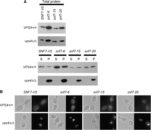
Figure 6.—
(A) Snf7 localization remains normal in alanine-scanning snf7 mutants during cell fractionation. Mid-log cultures were gently lysed and separated by centrifugation to generate a cytoplasm-containing supernatant (S) and an organelle-containing pellet (P). Fifty microliters of each fraction was run on 10% SDS–PAGES. Blots were probed with anti-V5-HRP antibody. (B) Snf7 localization remains normal in alanine-scanning snf7 mutants during immunofluorescence. Strains were grown to mid-log phase in M199 (pH 8) medium, fixed with 4% formaldehyde, spheroplasted, and attached to polylysine-treated wells for immunofluorescence. Samples were treated with anti-V5 antibody, followed by anti-mouse-IgG-alexafluor 488 (green).

