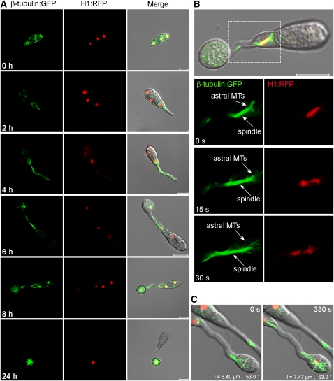
Figure 1.
Live-Cell Imaging of Microtubule Dynamics and Nuclear Division in M. oryzae during Germination and Appressorium Development.
(A) Time series of micrographs showing nuclear division during appressorium development in M. oryzae. The grg(p):H1:RFP and ccg1(p):bm1:sGFP gene fusion vectors were introduced into the M. oryzae wild-type strain Guy-11. Nuclei (red) and microtubules (green) were observed during conidial germination and appressorium development in M. oryzae.
(B) Astral microtubules were observed during mitotic spindle elongation and the concurrent rapid separation of chromosomes. MTs, microtubules.
(C) Continued expansion in diameter of the incipient appressorium during mitotic division. Bars = 10 μm.

