Figure 3.
PAS1 Is Required for Cell Patterning and Polarity in the Embryo Apex.
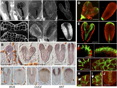
(A) Development of wild-type (top row) embryo at the dermatogen, globular, heart, torpedo, and late torpedo stage, respectively (from left to right). The first phenotypic alteration in pas1-3 embryos is visible at the heart stage with the absence of cotyledon formation (bottom row). Mutant embryos (bottom) were taken at the same respective stage (i.e., from the same silique) as the wild type (top row). Apical cells of pas1 embryos have lost their polar growth (inset, bottom) compared with the wild type (inset, top). Embryos were fixed and stained with propidium iodide.
(B) In situ hybridization of PAS1 mRNA during embryo development in the wild type. Embryos were taken at globular, young heart, late heart, young torpedo, and late torpedo stages (left to right).
(C) In situ hybridization of WUS, CUC2, and ANTEGUMENTA (ANT) mRNA in wild-type (left panel of each pair) and pas1-3 (right panel of each pair) embryos. Bars = 40 μm except for the inset in (A), which is10 μm.
(D) pDR5-GFP distribution in the wild type (left) and pas1-3 mutant (right).
(E) pPIN1:PIN1-GFP distribution in the wild type (left) and pas1-3 mutant (right).
(F) Detail of pPIN1:PIN1-GFP distribution in the tip of a wild-type cotyledon (left) and the apex of pas1-3 embryo (right) at heart stage.
(G) to (J) Immunolocalization of PIN1 in wild-type and pas1-3 embryos.
(G) Detail of PIN1 distribution in the tip of a wild-type cotyledon (left) and in the apex of the pas1-3 mutant where aggregates are visible (right).
(H) and (I) Altered polar distribution of PIN1 in the apex of the pas1-3 mutant with PIN1 localizations facing each other in adjacent cells ([H], arrow) or facing outward ([I], arrow).
(J) PIN1 polarity is normal in pas1-3 provascular and root pole cells.
Bars = 40 μm in (D) and (E), 20 μm in (F), 10 μm in (G) to (I), and 30 μm in (J).

