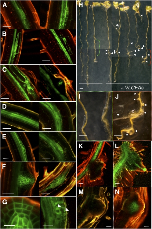
Figure 4.
VLCFAs Are Involved in Lateral Root Development and Auxin Polar Distribution.
(A) to (G) pDR5:GFP ([A] to [C]) and pPIN1:PIN1-GFP ([D] to [G]) expression during sequential steps of lateral root development in the wild type (left) and the pas1-3 mutant (right). In the pas1 mutant, PIN1-GFP was found accumulated inside primordia cells ([E] to [G], right) often in aggregates ([G], arrows).
(H) to (J) Exogenous application of VLCFAs restored lateral root development in pas1-3 mutants. Seedlings were grown in presence ([H], right) or absence ([H], left) of 200 μM fatty acids (18:0, 20:0, 22:0, and 24:0). Details of control ([I], green bracket in [H]) or treated roots ([J], red bracket in [H]) are shown. Arrows point to lateral root outgrowth in the pas1-3 mutant ([H] and [J]).
(K) to (N) VLCFA application restores polar auxin transport in pas1-3 lateral roots. Normal pDR5:GFP ([K] and [L]) and pPIN1:PIN1-GFP ([M] and [N]) expression patterns were observed in treated pas1-3 lateral root tips ([L] and [N]) but not in untreated mutant roots ([K] and [M]).
Bars = 45 μm in (A) to (F), 30 μm in (G) (left), 20 μm in (G) (right), 1 mm in (H), 300 μm in (I) and (J), and 20 μm in (K) to (N).

