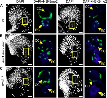
Figure 5.
Effect of CMT3 and DML Loss of Function on H3K9me2 in the Ovule.
(A) to (C) Ovules after fusion of the polar nuclei, as in Figure 3A, counterstained with DAPI (white). Left: Single optical sections; right: close-ups showing DAPI (blue) and H3K9me2 (green) overlays; projections of consecutive optical sections. Patterns shown below were observed in at least 30 ovules of each wild-type or mutant line. Two examples for each line are shown. EC, egg cell; CC, central cell. Bars = 10 μm.
(A) Wild-type ovule.
(B) dml1 dml2 dml3 mutant ovule showing an egg cell-like pattern of H3K9me2 in the central cell nucleus.
(C) cmt3 mutant ovule showing altered H3K9me2 signal specifically in the egg cell nucleus.

