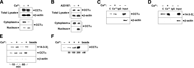
Figure 3.
Ca2+ stimulates CCTα binding to 14-3-3ζ. A, B) MLE cells were incubated in serum-free medium with (+) or without (−) calcium chloride (0.4 mM) for 1 h (A) or A23187 (100 nM) for 10 min (B). Cell lysates were harvested, subcellular fractions were isolated, and fractions were processed for CCTα immunoblotting. C, D) Coimmunoprecipitation. Cell lysates were harvested and incubated with primary antibody to CCTα or 14-3-3ζ for 1.5 h. Normal rabbit IgG was used as a negative control. Cell lysates were rotated overnight with anti-rabbit IgG beads at 4°C. The next day, immunoprecipitates were released from beads by 5 min of boiling in Laemmli buffer. Proteins were resolved by SDS-PAGE, followed by immunoblot analysis with CCTα (C) or 14-3-3ζ (D) and β-actin antibodies. E) His pulldown analysis. Cells were transfected with His-CCTα; after 24 h, cells were incubated in serum-free medium with (+) or without (−) calcium chloride (0.4 mM) for 1 h prior to harvest. Cellular lysates were purified on cobalt, and eluants processed for immunoblot analysis with 14-3-3ζ (above) or CCTα (below) antibodies, or β-actin antibodies to demonstrate the specificity of 14-3-3ζ-CCTα interaction. F) Purified CCTα was incubated with (+) or without (−) Ca2+ (0–200 nM) for 1 h, followed by 14-3-3ζ pulldown assays prior to CCTα immunoblotting. Data represent n = 3 separate experiments.

