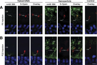
Figure 3.
MOP-NMP nanoparticle injection drives gene expression in cone photoreceptors. Double immunolabeling for transferred RDS (3B6, green) and cone OSs (S-opsin, red) with nuclear counterstain (DAPI, blue) was performed at PI-30 from eyes injected at P5 (A) or P22 (B). Images are single planes from a spinning disk confocal image stack. Representative cones from nanoparticle-injected animals are shown for each treatment. Cones in eyes injected with nanoparticles express transgenic NMP. Naked DNA-injected eyes express no transgenic NMP. Controls (right) are from saline-injected (A) and uninjected (B) animals. n = 3–5 animals/group. ROS, segment; COS, cone outer segment. Scale bar = 5 μm.

