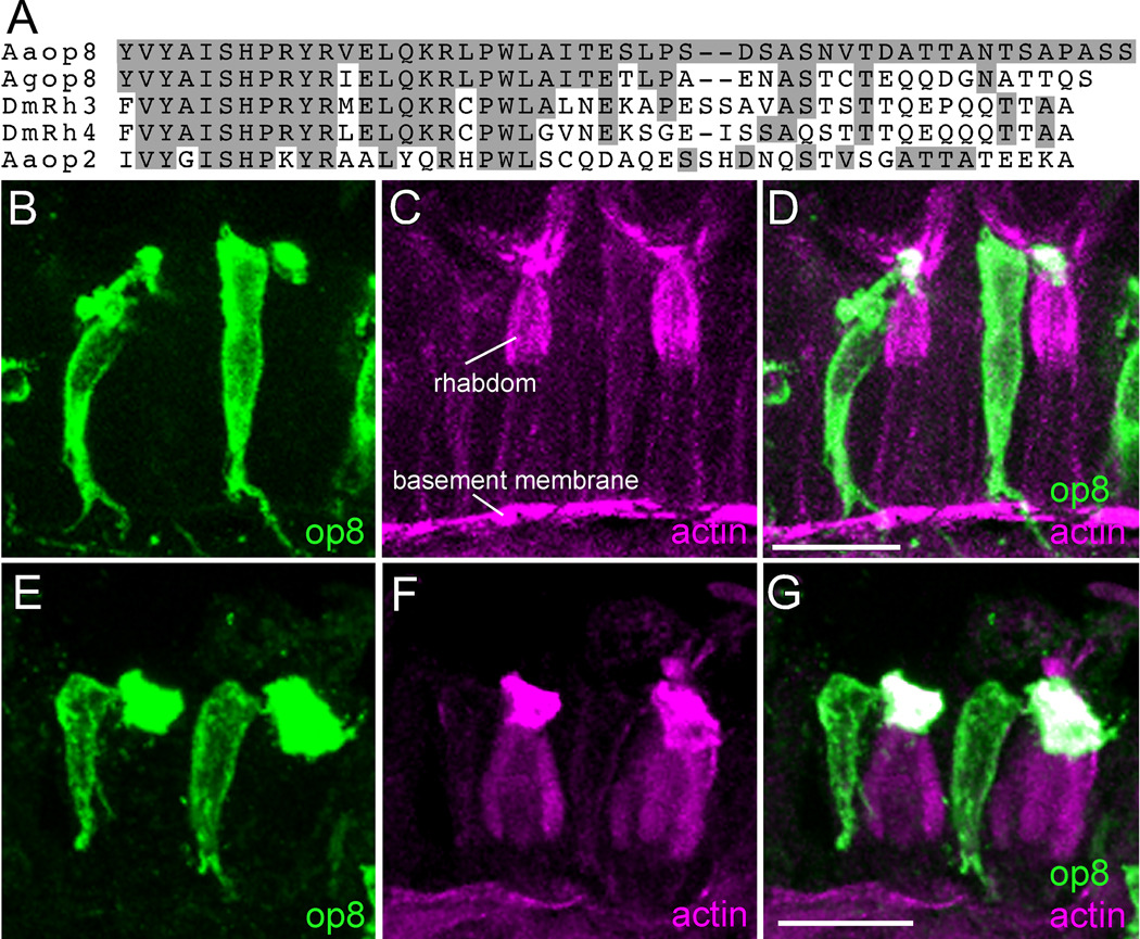
Figure 2.
Expression of op8 rhodopsin in Ae. aegypti and An. gambiae R7 photoreceptor cells. A: Multiple sequence alignment of the C-terminal regions of Ae. aegypti Aaop8 and Aaop2 with An. gambiae Agop8 and D. melanogaster Rh3 and Rh4 rhodopsins. Amino acids identical to the Aaop8 sequence are shaded gray. B, C, D: Ae. aegypti ommatidia labeled with Aaop8 antibody (green, B), phalloidin (magenta, C), and the resulting merged image (D). Phalloidin detects actin and heavily stains the rhabdom (labeled). E, F, G: An. gambiae ommatidia labeled with Aaop8 antibody (green, E), phalloidin (magenta, F), and the merged image (G). Scale bars in D and G are approximately 15 µm.

