
Fig. 2.
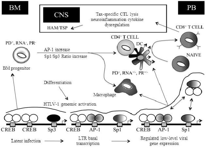
Overview of the HTLV-1 LTR regulation as it relates to HAM/TSP pathogenesis. The progressive stages of HAM/TSP are characterized by the presence of activated CD4+ and CD8+ T cells and macrophages in demyelinating lesions. At these sites an array of proinflammatory cytokines are produced facilitating further recruitment of inflammatory cells into the CNS. CD4+ T cells represent the chief source of viral gene expression and along with the help of antigen presenting cells such as dendritic cells activate CD8+ T cells. HTLV-1-specific CD8+ T cells traffic to and accumulate within the CNS throughout the course of neurologic disease. Therefore, the presence of activated HTLV-1-specific CD8+ CTL and macrophage populations in the CNS may result in the maintenance of a persistent CTL response against infected cells expressing viral antigens and proinflammatory cytokine-mediated bystander damage. During this process, differentiation of infected bone marrow progenitor cells lead to HTLV-1 genomic activation and an increased viral gene expression mediated by Tax-CREB dimer formation and the cell type-specific activity of different transcription factors such as AP-1, C/EBP and Sp1/Sp3. BM, bone marrow; CNS; central nervous system; PB, peripheral blood; PD, proviral DNA; PR, proviral replication; DC, dendritic cell