
Figure 1. Lymph heart myoblasts express skeletal muscle markers and not cardiac markers.
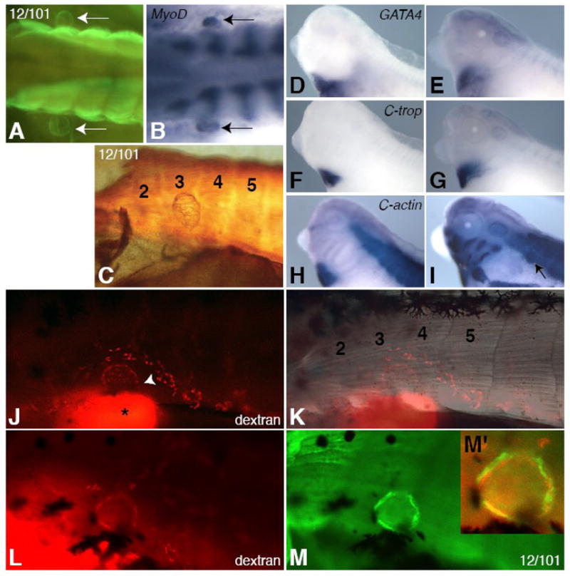
The 12/101 antibody marks lymph hearts in stage 42 tadpoles (A, C, M) and MyoD is expressed in differentiating lymph hearts at stage 40 (B, arrows). Two bilaterally symmetrical lymph hearts can be seen adjacent to the somites (A, B, arrows). The lymph heart forms in a region adjacent to somites 3 and 4 (C, K, trunk somites are numbered). Lymph heart muscle consists of a meshwork of myotubes (C). The lymph heart does not express cardiac markers GATA4 (D, E), cardiac troponin (F, G), or cardiac actin (H, I) at st. 33/34 (D, F, H) or st. 40 (E, G, I), except for cardiac actin at st. 40 (I, arrow), which marks all immature muscle (note expression in paraxial and head mesoderm in H, I). Rhodamine-dextran (mini-Ruby) injected subcutaneously into the posterior ventral tail fin drains anteriorly into the kidney (J, bright spot with asterisk) and into the lymph heart through vessels (J, arrowhead). The labeled lymph heart is located between somites 3 and 4 (K). Embryos injected with rhodamine-dextran (L, red) and then stained for the skeletal muscle antibody 12/101 (M, green) show co-expression (M′, yellow). All lateral views, anterior left, except for (A, B), which are dorsal views.