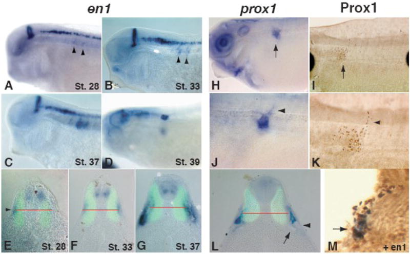
Figure 2. en1 and prox1 label the developing lymph heart musculature and endothelial tissue, respectively.

The expression of en1 is localized to the mid-hindbrain boundary, spinal interneurons, and anterior somites. The onset of somitic expression occurs at stage 28 (A) in a superficial region on a horizontal plane with the notochord (E, arrowhead, middle of notochord indicated by red line). The expression does not overlap with 12/101 staining in the differentiated muscle (green). Lateral views show early expression in the anterior somites (A–B, arrowheads), which intensifies and moves ventrally to occupy the final position of the lymph heart (C–D, G). A transverse section of a stage 33 tadpole (F) shows the intensity of expression increasing. By stage 37 (G), en1-positive cells have moved ventrally relative to the position of the notochord (red line) and are found directly above the glomus (arrow) and pronephric tubules (arrowhead). At stage 40, prox1 RNA (blue, H, J, L) and Prox1 protein (brown, I, K, M) are localized to the developing endothelial tissue of the lymph heart (H, I, arrows). (J, K) Higher magnification views of H and I illustrate the budding lymphatic vasculature dorsal to the lymph heart (arrowheads). (L) The expression of prox1 is slightly ventral to the notochord (red line) and dorsal to the pronephroi (arrow) and does not overlap with differentiated muscle (12/101, green). (M) Prox1 (brown) and en1 (blue) expressing cells co-localize at the site of lymph heart formation (arrow). The epidermis has sloughed off during the processing of this embryo.
