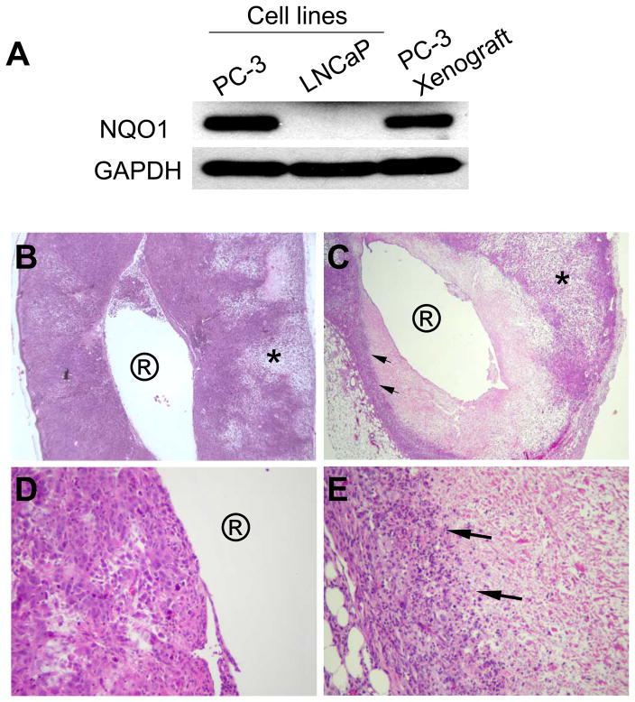
Fig. 5.
Histological examination (H&E) of PC-3 tumor xenograft sections after millirod implantation confirmed significant anti-tumor effects of β-lap in vivo. (A) Western analyses demonstrating NQO1 expression in explanted PC-3 xenografts. Cell lysate of parental LNCaP cells (NQO1−) was used as negative control. (B) A cross section of tumors treated with HPβ-CD-loaded millirod (4× magnification). Circled ‘R’ represents the millirod implantation site, while * indicates areas of patchy necrosis. (C) A cross section (4× magnification) of a tumor xenografts 6 days after implantation of β-lap-loaded millirods. (D) and (E) represent increased magnifications (40×) of (B) and (C), respectively.

