
Figure 4. Proposed working model of the human RLC.
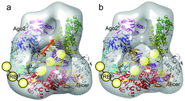
The 3D density map of RLC is shown as a semi-transparent iso-surface. The atomic model of the DExH/D domain (red ribbon), the Giardia Dicer atomic model (gray-yellow-green-orange ribbon, color coding the same as in Figure 1b), and the Thermus thermophilus Argonaute (gray-cyan-orange-pink-blue, color coding the same as in Figure 3b) are docked in the density map. TRBP is illustrated as a string of three yellow spheres with a flexible linker connecting it to the DExH/D domain. Its motion range, based on our experimental results, is marked by the dashed yellow arrows. In (a), an atomic model of the siRNA (paired spirals with the guide strand in purple color and the passenger strand in yellow color) is aligned vertically between the Dicer's RNase III domain flat surface and Ago2's PAZ domain. This panel illustrates the state proposed in our model for the dicing of dsRNA by Dicer. In this state the PAZ domain of Ago2 could engage the newly diced end of the siRNA, as illustrated by the red arrow. In (b), the distance between the PAZ domains of Ago2 and Dicer allows a perfect accommodation of the 22 nt siRNA between them. This state of Ago2 could be stabilized by interactions with TRBP. Thus, this panel illustrates the hypothetical state after transfer of the newly diced siRNA's onto Ago2's PAZ domain while the other end remains bound to Dicer's PAZ domain. The flexible TRBP could help the transfer efficiency and correctness.