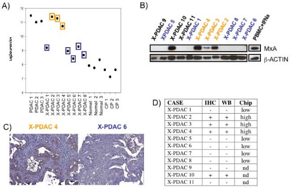
Figure 3.
MxA protein expression in xenografted primary pancreatic adenocarcinomas. A) MxA expression level in microarray data analysis expressed as log2 ratio; orange and blue colors represent higher and lower expression transcript, respectively. B) Western Blot analysis of MxA in 11 xenografted primary pancreatic adenocarcinomas (X-PDAC). C) Example of MxA immuno positive (X-PDAC 4) and MxA immuno negative (X-PDAC 6) samples. D) Correlation of MxA immunohistochemistry, Western Blot and microarray data.

