Figure 4.
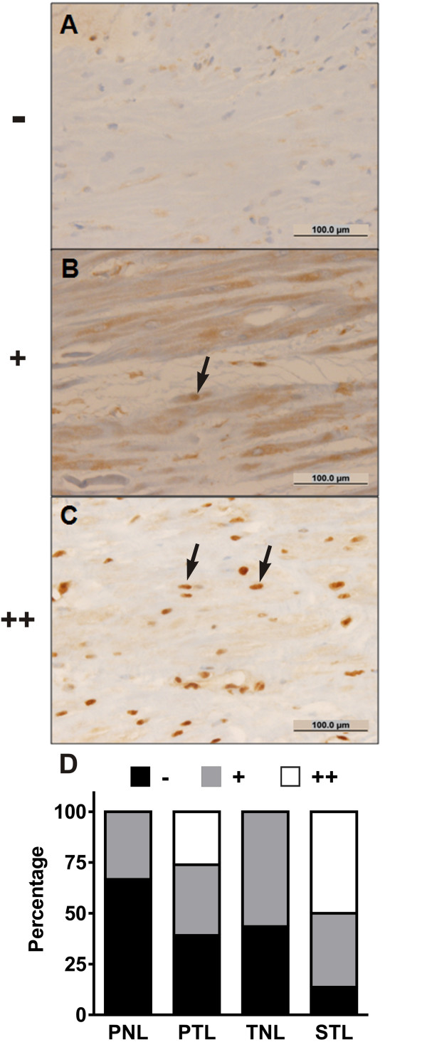
Immunohistochemical localization of NF-κB p65 in term and preterm myometrium. (A-C) Representative photomicrographs demonstrating the graded scale used to semi-quantitatively assess the extent of p65 immunolabeling in myocytes: (A) no nuclear p65 immunoreactivity (-); (B) isolated nuclear p65 immunoreactivity in a minority of myometrial cells (+); (C) diffuse nuclear p65 immunoreactivity in a majority of myometrial cells (++). The arrows indicate typical myocytes with nuclear p65 immunolabeling. Bars = 100 μm. (D) Percentage of cases with absent, isolated, and diffuse nuclear p65 staining in myometrial myocytes from biopsy specimens obtained from the following cohorts: preterm no labor (PNL, N = 21), spontaneous preterm labor (PTL, N = 21), term no labor (TNL, N = 23), and spontaneous term labor (STL, N = 21). There were significantly greater proportions of cases exhibiting diffuse nuclear p65 immunolabeling in the PTL and STL groups relative to the unlabored cohorts (P < 0.001, Chi-square test).

