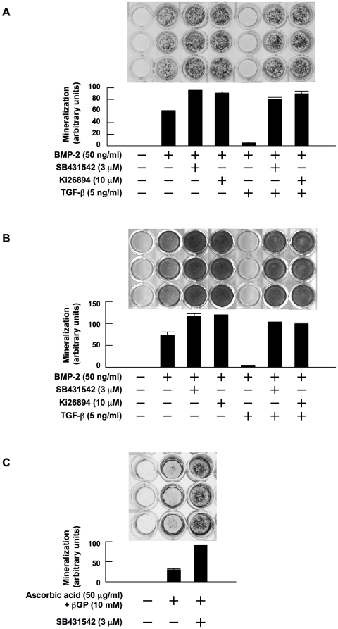
Figure 1. TGF-β suppresses and TGF-β inhibitors enhance OB differentiation.
MC3T3-E1 cells (A) and primary bone marrow stromal cells (B) were cultured for 14 and 28 days, respectively, in 24-well culture plates in α-MEM containing 10% FBS supplemented with β-glycerophosphate and ascorbic acid (osteogenic medium). rhBMP-2, SB431542, Ki26894 and rhTGF-β were added at 50 ng/mL, 3 µM, 10 µM and 5 ng/mL to the indicated wells, respectively. After culturing, mineralized nodules were visualized by von Kossa staining. C. MC3T3-E1 cells were cultured for 21 days in osteogenic media in the absence of BMP-2. SB431542 was added at 3 µM to the indicated wells. Mineralized nodules were visualized by von Kossa staining. The data with mineralized nodule formation were quantified by densitometric analyses.

