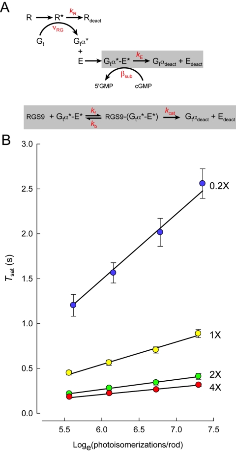
Figure 2.
Michaelis-module for the RGS9-dependence of Gtα-PDE* deactivation reveals the rate constants of RGS9 binding and catalysis, and constrains R* lifetime. (A) Standard scheme for phototransduction, in which the Michaelis module for RGS9-mediated GTP hydrolysis (gray box below) was substituted for the first order decay of Gtα-PDE* (gray box above). (B) The time that flash responses remained in saturation (Tsat) as a function of the natural log of the number of R* (photoisomerizations) produced by each flash for mouse rods expressing a 20-fold range of RGS9 complex.54 Error bars represent SEMs. Straight lines are the best-fitting curves produced using simplex searches of the solutions to the differential equations representing the expanded scheme in (A). Parameter values were kR = 33 seconds−1, kf = 0.051 μm2 s−1, kb = 13.8 seconds−1 and kcat = 52.8 seconds−1. Reprinted with permission from Burns ME, Pugh EN Jr. RGS9 concentration matters in rod phototransduction. Biophys J. 2009;97:1538–1547. © 2009 Biophysical Society Published by Elsevier Inc.

