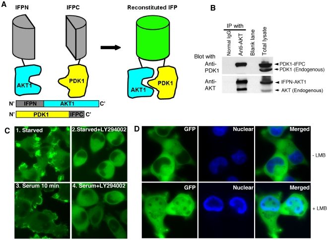
Figure 1. AKT1 interaction with PDK1 stabilized by the reconstituted IFP.
(A) Schematic diagram showing the split IFP reconstitution mediated by the interaction between AKT1 and PDK1. IFPN was fused to the N-terminus of AKT1. IFPC was fused to the C-terminus of PDK1. Neither fusion protein was fluorescent. The interaction between AKT1 and PDK1 brings IFPN and IFPC in proximity and reconstitutes IFP fluorescence. The reconstituted IFP stabilizes the PDK1-IFPC::IFPN-AKT1 complex. (B) Association of PDK1-IFPC with IFPN-AKT1. HeLa cells stably expressing the PDK1-IFPC::IFPN-AKT1 complex were lysed in NP40 lysis buffer. Immunoprecipitation was performed with the indicated antibodies (top labels), followed by western blotting with anti-PDK1 (upper blot) and anti-AKT (lower blot). Lane 1, IP with normal IgG. Lane 2, IP with anti-AKT1. Lane 3, blank. Lane 4, total cell lysate. (C) Translocation of the PDK1-IFPC::IFPN-AKT1 complex upon PI3K signaling activation or inhibition. Cells stably expressing the PDK1-IFPC::IFPN-AKT1 complex were serum starved for overnight then treated with LY294002 (20 µM) for 3 hours (image 2), 10% serum for 10 minutes (image 3), and LY294002 (20 µM) for 3 hours and then 10% serum for 10 minutes (image 4). At least 100 cells were examined from different fields for each sample with 100% of the examined cells in each sample showing the same fluorescence localization presented. (D) Accumulation of the PDK1-IFPC::IFPN-AKT1 complex in nucleus with LMB treatment. HeLa cells stably expressing the PDK1-IFPC::IFPN-AKT1 complex were starved overnight and then treated with LMB at 50 nM for 3 hours. Nuclei were stained with Hoechst 33342.

