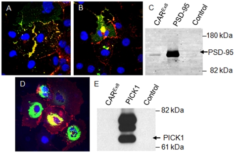
Figure 3. hCAREx8 co-localizes and interacts with PSD-95 but not PICK1.
Panel A shows co-localization (yellow) of hCAREx8 (red) and PSD-95-GFP (green). In contrast, in panel B, hCAREx8-PDZ does not co-localize at the junctions of cells. hCAREx8-PDZ localizes to the junctions between cells whereas PSD-95-GFP fluorescence remains diffuse. Panel C shows immunoprecipitation of PSD-95-GFP with the hCAR specific extracellular domain monoclonal antibody RmcB, GFP antibody, but not a control antibody (MopC). Panels D and E shows the lack of co-localization and immunoprecipitation between hCAREx8 (junctional) and PICK1-GFP (perinuclear). Confocal microscopy (60x oil immersion).

