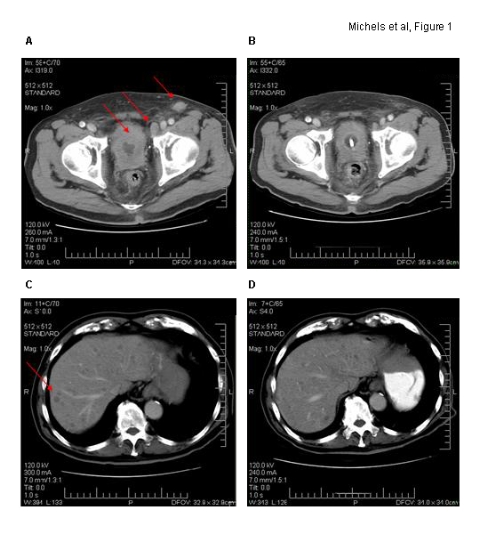
Fig. 1.

Axial computer tomographic images. Corresponding imaging sections before (A/C) and at end (B/D) of capecitabine chemotherapy are demonstrated. Marked bladder wall thickening with significant pelvic/inguinal lymphadenopathy and multiple liver metastases are noted (red arrows) on baseline computer tomography.
