Figure 1.
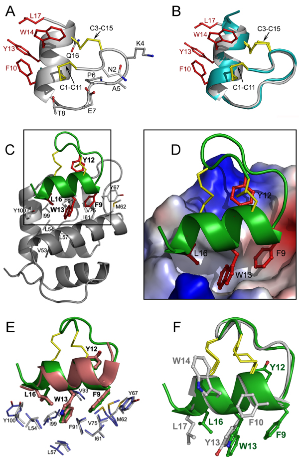
Crystal structures of stingin-5 and of stingin-1 in complex with MDM2. (A) Ribbon representation of the overall structure of stingin-5. All side chains are shown as ball-sticks. Residues grafted to the apamin sequence are shown in red and disulfide bonds in yellow. (B) Superposition of the backbones of stingin-5 (grey) and apamin (cyan). (C) The co-crystal structure of synthetic 25–109MDM2 (grey) and stingin-1 (green). Side chains of Phe9, Tyr12, Trp13, and Leu16 of stingin-1 are colored in red, and residues of MDM2 shaping the stingin-binding pocket shown as grey sticks. (D) Close-up view of the interface of the protein-peptide complex. The electrostatic potential at the molecular surface of MDM2 is displayed as negative in red, positive in blue, and apolar in white. (E) Stingin-1 (green) and PMI (pink), shown in a ribbon and stick representation, from their respective complexes with (superimposed) synMDM2. Residues shown as thin sticks line the hydrophobic cavity of MDM2 - blue residues from MDM2 complexed with PMI, and grey residues from MDM2 complexed with stingin-1. (F) Superimposed stingin-1 (green) and stingin-5 (grey).

