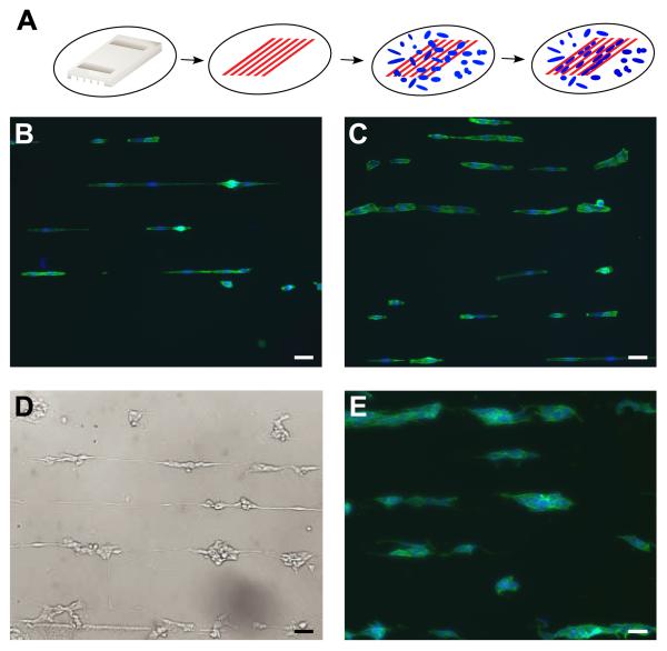
Figure 3. Patterning NBL cells with F108.
A. PDMS stamp is placed in contact with a 60 mm tissue culture dish and F108 (red) is flowed through the channels. Cells (blue) land at random, detect F108 stripes and migrate to stripes of more adhesive tissue culture plastic. SH-EP cells complied to narrow (B) and wide (C) stripes of tissue culture plastic separated by stripes of F108. Both N-type cell lines, SH-SY5Y (D) and IMR-32 (E), are also patterned effectively by F108. Cells were stained with phalloidin (green) and bisbenzimide (blue). Scale bar = 50μm.

