Figure 1.

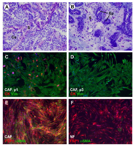
Characterization of fibroblasts isolated from human breast tissue samples: A) Hematoxylin-eosin-stained frozen section of tissue piece taken from infiltrating ductal carcinoma sample prior to tissue mincing. B) Hematoxylin-eosin-stained frozen section of tissue piece taken from adjacent normal mammary tissue sample prior to mincing. C) Carcinoma-associated fibroblasts (CAF) from human breast carcinoma sample at passage 1 (p1). The cells were immunofluorescence-labeled for epithelial marker cytokeratin (red signal) and the mesenchymal marker vimentin (green signal). Note a few epithelial cells and epithelial cell fragments among the fibroblasts. D) Carcinoma-associated fibroblasts (CAF) from the same human breast carcinoma sample as shown in panel “C” at passage 3 (p3). The cells were immunofluorescently labeled for epithelial marker cytokeratin (red signal) and mesenchymal marker vimentin (green signal). At this stage, no epithelial cells are left. E) CAF labeled for FSP1 (red signal) and αSMA (green signal). F) Normal fibroblasts (NF) labeled for FSP1 (red signal) and αSMA (green signal). Abbreviations: Ca = carcinoma; S = stroma). Original magnification: 200× for all images. Blue signal in panels C – F: nuclear label DAPI
